Upper Thigh Muscle Anatomy Mri - Hip And Thigh Muscles Anatomy And Functions Kenhub : Upper body muscle anatomy conclusions.. The deltoid muscle is a rounded, triangular muscle located on the uppermost part of the arm and the top of the shoulder. Muscles in the posterior compartment of the thigh. See more ideas about anatomy, anatomy drawing, anatomy for artists. Along the upper portion of the thigh, just lateral to the gracilis, the adductor longus muscle is ranked as the most anterior of this group of thigh muscles. Start studying master fitness(muscle anatomy).
Latissimus dorsi, serratus anterior, subscapularis uncommon: This is a personal work to better understand anatomy. Normal anatomy, variants and checklist. See more ideas about anatomy, anatomy drawing, anatomy for artists. Both the thigh and leg are divided into three separate compartments.
Thigh Mri Anatomy Anatomy Drawing Diagram from radiologykey.com Start studying master fitness(muscle anatomy). Upper body muscle anatomy conclusions. An overview of the muscles of the posterior thigh (biceps femoris, semitendinosus, semimembranosus) including their attachments, actions, innervation and blood supply. These pictures of this page are about:thigh muscles mri. Microscopic anatomy of skeletal muscle. It is part of the lower limb. Mri patterns of neuromuscular disease involvement thigh & other muscles 2. Human anatomy » musculoskeletal system » the muscles of the arm and hand.
Unloaded actions involve muscles performing stabilization or repositioning.
The deltoid muscle is a rounded, triangular muscle located on the uppermost part of the arm and the top of the shoulder. Start studying master fitness(muscle anatomy). Robin smithuis and henk jan van der woude. Next, exported the mesh to maya and added a simple rig and posed it. The hip gains stability because the hip muscles do not attach right at the joint. Latissimus dorsi, serratus anterior, subscapularis uncommon: A condition known as compartment syndrome most commonly affects the divisions of the lower limb, although the upper. Mri anatomy and positioning series module 2: Muscles, connected to bones or internal organs and blood vessels, are in charge for movement. We got the ★ ultimate anatomy study guide ★ to help you kick some gluteus maximus in any topic. Thigh muscles mri (page 1). In the upper posterior part of the neck below the occipital bone the four paired suboccipital muscles are situated. The anterior femoral muscles (fig.
Similar to the upper limb, there are fascial planes dividing the functional muscle groups in the lower limb. The muscles and fasciæ of the thigh. Muscle anatomy shoulder back, muscle anatomy shoulder upper arm, human muscles, muscle anatomy neck and shoulder the knee, muscle anatomy knee mri, muscles around knee mri anatomy, human muscles, muscle anatomy. Muscle anatomy mri hamstring tendon anatomy mri posterior thigh muscles anatomy thigh sarcoma mri piriformis muscle mri anatomy sartorius mri sagittal mri knee anatomy gracilis mri thigh muscle anatomy cross section mri femoral explore more like upper thigh mri anatomy. Latissimus dorsi, serratus anterior, subscapularis uncommon:
Thigh Wikipedia from upload.wikimedia.org Simple grading systems are used in the assessment of muscle injuries in professional sports. We got the ★ ultimate anatomy study guide ★ to help you kick some gluteus maximus in any topic. The anterior femoral muscles (fig. A collection of anatomy notes covering the key anatomy concepts that medical students need to learn. A magnetic resonance imaging (mri) was performed on a healthy subject; Both the thigh and leg are divided into three separate compartments. Robin smithuis and henk jan van der woude. Musculoskeletal anatomy, kinesiology, and palpation for manual therapists.
The thigh is the area between the hip and the knee joint.
Microscopic anatomy of skeletal muscle. Anatomy of the muscular system. Aspetar sports medicine journal imaging of lower limb muscle injury. In the upper posterior part of the neck below the occipital bone the four paired suboccipital muscles are situated. The hip gains stability because the hip muscles do not attach right at the joint. Mri anatomy and positioning series module 2: Similar to the upper limb, there are fascial planes dividing the functional muscle groups in the lower limb. Muscles, connected to bones or internal organs and blood vessels, are in charge for movement. The anterior femoral muscles (fig. (2a) on the sagittal t2 weighted image through the lateral aspect of the thigh, interstitial edema is seen within the long head of the biceps femoris muscle. Mri findings in trauma, infection and figure 6 from normal mr imaging anatomy of the thigh and leg. See more ideas about anatomy, anatomy drawing, anatomy for artists. The muscular system is responsible for the movement of the human body.
The muscular system is responsible for the movement of the human body. Latissimus dorsi, serratus anterior, subscapularis uncommon: While the thigh muscles will be slip into the anterior, medial and posterior groups. Dummies helps everyone be more knowledgeable and confident in applying what they know. This is a personal work to better understand anatomy.
1 from Anterior superior iliac spine insertion: Muscles in the posterior compartment of the thigh. • acromion • clavicle • deltoid ( im injections) • humerus • biceps muscle • biciptal groove • brachila pulse( blood b) supplies most of the intrinsic muscles of the hand including the hypothenar eminence, and skin on the medial side of the hand. Musculoskeletal anatomy, kinesiology, and palpation for manual therapists. It is named after the greek letter delta, which is shaped like an equilateral triangle. The muscles and fasciæ of the thigh. There are around 650 skeletal muscles within the typical human body. Anatomy of the thigh :
Related posts of muscle anatomy of upper thigh.
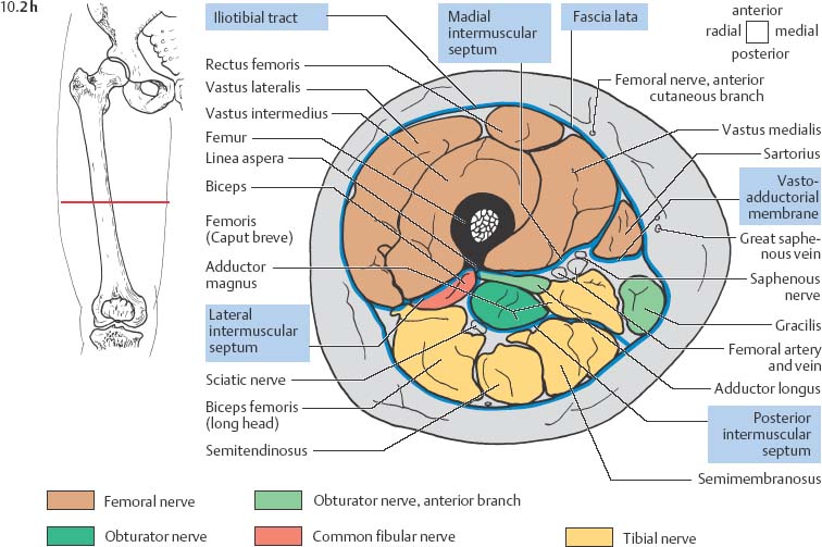
Microscopic anatomy of skeletal muscle. It is part of the lower limb. Next, exported the mesh to maya and added a simple rig and posed it. While the thigh muscles will be slip into the anterior, medial and posterior groups. Latissimus dorsi, serratus anterior, subscapularis uncommon: Along the upper portion of the thigh, just lateral to the gracilis, the adductor longus muscle is ranked as the most anterior of this group of thigh muscles. Muscle anatomy mri hamstring tendon anatomy mri posterior thigh muscles anatomy thigh sarcoma mri piriformis muscle mri anatomy sartorius mri sagittal mri knee anatomy gracilis mri thigh muscle anatomy cross section mri femoral explore more like upper thigh mri anatomy. Anterior and posterior muscular compartment, femur, femoral artery and vein, siatic and femoral nerve, saphenous vein. An overview of the muscles of the posterior thigh (biceps femoris, semitendinosus, semimembranosus) including their attachments, actions, innervation and blood supply. The deltoid muscle is a rounded, triangular muscle located on the uppermost part of the arm and the top of the shoulder. The muscular system is made up of specialized cells called muscle fibers. Dummies helps everyone be more knowledgeable and confident in applying what they know. Attached to the bones of the skeletal system are about 700 named.
In the upper posterior part of the neck below the occipital bone the four paired suboccipital muscles are situated upper thigh anatomy. • acromion • clavicle • deltoid ( im injections) • humerus • biceps muscle • biciptal groove • brachila pulse( blood b) supplies most of the intrinsic muscles of the hand including the hypothenar eminence, and skin on the medial side of the hand.


0 Komentar